Biopsy Site Markers
Tumark® - Identify with confidence
Markers from the Tumark® family are used for marking biopsy sites and suspicious lesions in the breast tissue. 18-gauge thin, sharp puncture cannulas enable precise marking and ensure minimally invasive treatment. The ergonomic handle design allows for single-handed use under ultrasound-guidance.
Due to their shapes, the markers anchor firmly in the tissue and are detectable in ultrasound, X-ray and MRI. All markers are made from the biocompatible implant material Nitinol and remain visible in the tissue.
- Intelligent 3D design of the markers provides long-term visibility in ultrasound, X-ray and MRI1
- Markers expand into shapes upon deployment and anchor firmly in the tissue1
- Ergonomic handles allow for single-handed use
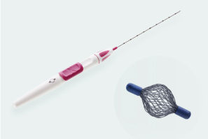
Tumark Vision
- 3D structure anchors firmly in the tissue
- Mesh structure made of 48 individual wires
- Mesh structure in a 3D spherical design leads to high echogenicity in ultrasound imaging regardless of transducer positions1
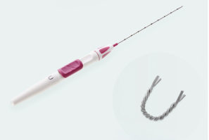
Tumark Professional / U
- Lateral anchor “feet” provide secure anchoring in the tissue1
- Marker material is sandblasted and twisted
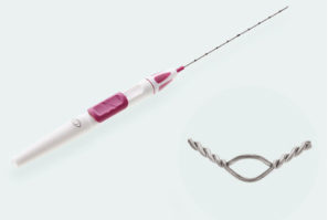
Tumark Eye
- Angled ends provide secure anchoring in the tissue1
- Bent shape with opening and twisted ends
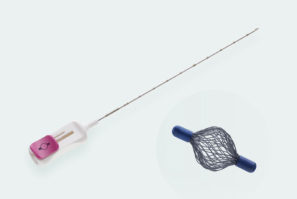
Tumark Vision MRI
- Tumark Vision marker with MRI-compatible deployment system
- System suitable for MRI-guided tissue marking up to 3 Tesla
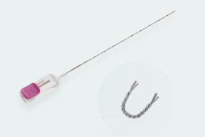
Tumark MRI
- U-shaped marker with MRI-compatible deployment system for MRI-guided operations up to 3 Tesla
- Compact construction of application system for use in the MRI gantry area
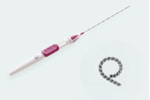
Tumark Professional Q
- Q-shape becomes firmly anchored in the tissue1
- Marker material is sandblasted and twisted
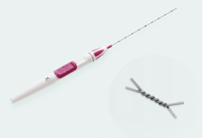
Tumark Professional X
- Lateral anchor “feet” provide secure anchoring in the tissue1
- Twisted marker with angled ends
 English (EUR)
 English
 Deutsch
 Español
 Italiano
 Français
 Nederlands
